Mapping the Fruit Fly’s Brain Reveals Cognitive Secrets
The digital reconstruction of the fruit fly brain represents a monumental leap in neurobiology, offering a mapping of neural architecture that could fundamentally alter how humanity understands and treats cognitive disorders.
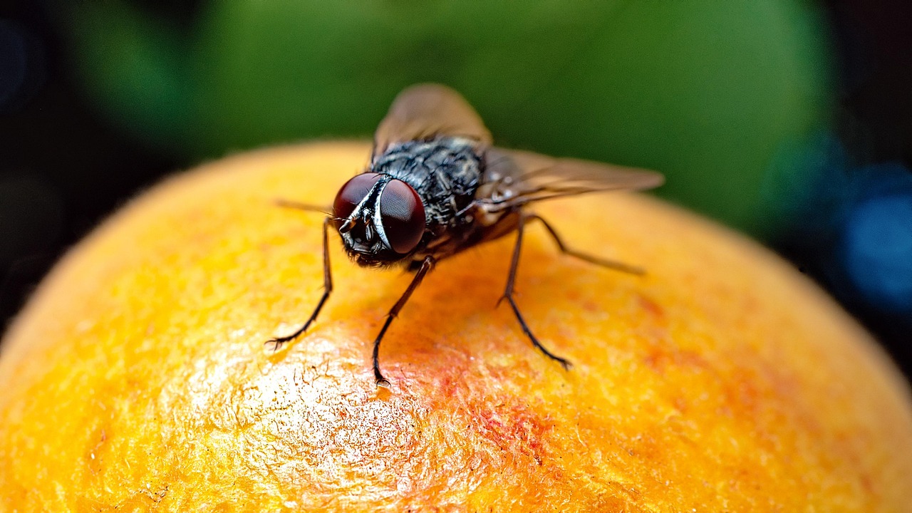
In the annals of neuroscience, few milestones rival the recent completion of the adult fruit fly connectome. Researchers have successfully mapped every neuron and synapse within the brain of Drosophila melanogaster, the common fruit fly. This poppy-seed-sized organ, despite its diminutive scale, contains a labyrinthine network of approximately 140,000 neurons and over 50 million synaptic connections. The achievement, spearheaded by the FlyWire Consortium-a massive global collaboration involving Princeton University, the University of Cambridge, and numerous other institutions-is not merely a triumph of biological cataloging. It represents a functional atlas that allows scientists to trace the flow of information through a complex living system, providing a blueprint that may eventually unlock the mysteries of the human mind and the devastating diseases that afflict it.
The Cartography of a Microscopic Masterpiece
Mapping a brain is a task of staggering complexity. To understand the scale of the fruit fly project, one must look at the history of connectomics. Previously, scientists had mapped the brain of the C. elegans worm, which possesses a mere 302 neurons, and the larval fruit fly, which has roughly 3,000. Jumping to the 140,000 neurons of an adult fruit fly represents a leap of several orders of magnitude. This project, documented in a suite of papers in the journal Nature, provides the most detailed wiring diagram of an adult animal brain ever created.
The process of creating this map was a monumental technological feat. It began with 21 million high-resolution images of a female fruit fly’s brain. From there, artificial intelligence models were employed to trace the winding paths of neurons through the three-dimensional space. However, AI alone was not sufficient; the intricate “branches” and “twigs” of neurons required human verification. This led to a unique “team science” approach where professional neuroscientists collaborated with citizen scientists and online gamers. Over 33 person-years were dedicated to proofreading and annotating the results. Sebastian Seung, a professor of neuroscience and computer science at Princeton, noted that without the assistance of AI, the project would have required nearly 50,000 person-years to complete.
The resulting map, known as the connectome, is more than a static image. It is a searchable database. Through tools like the FlyWire Codex-described as “Google for the connectome”-researchers can now navigate specific neural pathways as easily as one might navigate a city using digital maps. This atlas includes annotations for cell types, neurotransmitter profiles, and the specific regions where signals are processed, such as the subesophageal zone (SEZ), which interacts with almost every part of the brain.
Synopsis
Researchers have created a detailed connectome of the adult fruit fly brain, tracing nearly 140,000 neurons and more than 50 million synapses. The open atlas reveals how circuits connect perception, memory, decision-making, and action, while giving scientists a manageable system for studying disease, efficient computation, and the neural principles that may extend to larger brains.
Why the Fruit Fly is a Surrogate for the Human Mind
A common question arises: why spend years mapping the brain of an insect that most people consider a household pest? The answer lies in the deep evolutionary ties between flies and humans. Despite 500 million years of divergent evolution, fruit flies share approximately 60 percent of human DNA. More importantly, three out of four human genetic diseases have a parallel in the fruit fly.
Fruit flies exhibit surprisingly sophisticated behaviors that mirror human cognition. They form long-term memories, engage in complex social interactions, navigate over vast distances using visual cues, and even experience sleep cycles and circadian rhythms. By mapping the circuits that govern these behaviors in a fly, researchers can hypothesize about similar structures in the human brain. The human brain contains roughly 86 billion neurons and 100 trillion synapses, making it far too complex to map in its entirety with current technology. The fruit fly serves as a “Goldilocks” organism-complex enough to exhibit meaningful behavior, yet small enough to be fully mapped.
Understanding how a fly’s brain processes a scent or decides to move toward a food source provides a fundamental understanding of “biological logic.” When a brain is healthy, the wiring follows specific patterns. In diseases like Alzheimer’s, Parkinson’s, or dementia, these patterns are disrupted. The fly connectome provides a “control” map, allowing scientists to simulate damage and test how neural circuitry might be repaired. By understanding the “street names and buildings” of the fly brain, researchers are better equipped to identify where things go wrong in the human version.
From Biology to Silicon: The Rise of Fly-Powered AI
The open-source release of the fly connectome has triggered a wave of creative and scientific experiments that bridge the gap between biology and artificial intelligence. Because the map is digital, it can be imported into computer models, essentially “uploading” the logic of a fly’s brain into a virtual environment. This has led to the development of specialized AI models that do not rely on traditional large language model (LLM) architectures, but rather on the actual firing patterns of biological neurons.
One such experiment, dubbed “PitchFly,” involved training a digital representation of the fly brain on thousands of headlines from the technology publication WIRED. The goal was to see if the fly’s “remixing” of patterns could generate viable story ideas. While the output was often surreal-generating headlines like “Everyone Wants Cooking. Nobody Has Solved Donald Trump”-it demonstrated that even a tiny biological model can perform pattern recognition and creative synthesis.
Other developers have pushed this further. An engineer at Coinbase created “StonkFly,” an AI model that uses the fly connectome to make stock market trades. Others have trained the virtual fly brain to play video games like Doom, Minecraft, and Beat Saber, or to solve Rubik’s Cubes. While some of these projects are lighthearted, they serve as a proof of concept for “agentic AI.” They show that a specialized model based on biological intelligence can often be more efficient at specific tasks than a massive, energy-hungry LLM. Sometimes, a tiny brain is all that is needed to solve a specific problem.
Our Perspective
The fruit fly connectome matters less as a miniature version of the human brain than as a full-scale experiment in understanding a nervous system from the inside out. Its greatest value will come from pairing the static wiring diagram with genetics, physiology, behavior, and computation—turning a map of connections into an explanation of how living circuits adapt, fail, and produce intelligent action.
Mapping the Projectome and Information Flow
A significant discovery made during the FlyWire project was the detailed mapping of the “projectome”-the set of long-range connections that link different regions of the brain. Researchers found that the fruit fly’s brain is highly symmetrical but possesses specific “hub” neurons that facilitate communication across the hemispheres.
The study highlighted the role of the subesophageal zone (SEZ), a region that had been poorly understood in previous research. The new connectome revealed that the SEZ acts as a primary hub for sensory input and motor output. It receives a massive portion of the signals entering the brain and sends out almost all the signals to the motor neurons that control the fly’s legs and wings. This makes the SEZ the “central station” of the fly’s behavioral responses.
Additionally, researchers traced the ocellar circuit, which processes light through the fly’s simple eyes to help it orient its body during flight. By following the “wires” from the eyes to the motor centers, scientists can now see exactly how a visual stimulus is converted into a physical movement. This level of granularity is what neuroscientists have dreamed of for decades. It allows them to “close the loop” between a specific neuron and a specific behavior.
The Ethical and Philosophical Frontiers
As the ability to simulate biological brains grows, so do the philosophical questions surrounding the technology. When the firing patterns of 140,000 neurons are perfectly replicated in a digital space, is there a form of consciousness emerging? While most scientists agree that a fruit fly-and certainly a digital simulation of one-lacks the self-awareness associated with higher mammals, the replication of its neural architecture sparks debates about the ethics of brain mapping.
Software engineer Alex Wormuth, who worked with the connectome, noted that the project “sparks questions about ethics and whether there is any consciousness in the replication of a fly’s brain.” As the science moves toward mapping larger animals, such as mice or even monkeys, these questions will become more pressing. For now, the fly serves as a safe and invaluable testing ground for these ideas, allowing humanity to explore the mechanics of thought without the profound ethical weight of simulating a human-like consciousness.
Glossary
- Connectome — A wiring diagram that describes the connections among neurons in a nervous system.
- Drosophila melanogaster — The common fruit fly used extensively in genetics and neuroscience research.
- Neural circuit — A connected group of neurons that processes information or helps control behavior.
- Neuron — A nerve cell that receives, processes, and transmits signals.
- Synapse — A junction where one neuron communicates with another cell.
- Synaptic connection — A functional communication link formed at a synapse.
- Subesophageal zone — A major fly-brain region that integrates signals and sends extensive outputs to motor systems.
- Cell type — A class of neurons grouped by shared structure, molecular features, or connectivity.
- Neural modulation — Chemical or physiological changes that alter how neural circuits respond.
- Connectomics — The study of large-scale neural connectivity using maps such as connectomes.
A Forerunner to Human Treatments
The ultimate goal of the FlyWire project and the NIH’s BRAIN Initiative is to apply these lessons to human health. John Ngai, the director of the NIH’s BRAIN Initiative, described the fly connectome as a “forerunner” to mapping the connections of larger mammalian brains. By establishing the methodology for mapping and proofreading a brain of this scale, researchers have paved the way for more ambitious projects.
The potential for “tailored treatments” is the most promising outcome. If scientists can identify the exact circuit that governs a specific behavior or cognitive function, they can develop drugs or therapies that target those specific neurons rather than the entire brain. This could lead to more effective treatments for mental health disorders and neurodegenerative diseases with fewer side effects.
Furthermore, the fly connectome allows researchers to study how “damage” to the wiring diagram affects function. By digitally “breaking” certain synapses in the model, they can observe the resulting behavioral changes and then search for those same patterns in human patients. This “top-down” approach to neuroscience-moving from a complete map to specific disorders-is a radical departure from traditional “bottom-up” methods that focus on individual cells in isolation.
The Beginning of the Work, Not the End
The completion of the adult fruit fly connectome is not the conclusion of a scientific journey, but the beginning of a new era. As Sven Dorkenwald of the FlyWire Consortium stated, “I think the hard work is ahead. This is a beginning, not the end of the work.” With the atlas now in hand, the global scientific community can begin the painstaking process of testing hypotheses, running simulations, and uncovering the hidden logic of the brain.
The humble fruit fly, often dismissed as a nuisance, has provided humanity with its first clear look at the internal wiring of a complex mind. As researchers continue to explore this digital atlas, the secrets of memory, navigation, social interaction, and perhaps even the nature of consciousness itself may finally begin to reveal themselves. The poppy-seed-sized brain of Drosophila melanogaster has become the key to understanding the most complex structure in the known universe: the human brain.
Through the collaborative efforts of thousands of people and the power of artificial intelligence, the “poppy seed” has been cracked open, revealing a world of connections that link us all-fly and human alike-to the fundamental laws of biological intelligence. The secrets of the fruit fly are, in many ways, our own secrets, and the map to its mind is a map to our future health and understanding.
Frequently Asked Questions
What did researchers discover by mapping the fruit fly’s brain?
How was the fruit fly brain connectome created?
Why is the connectome important for understanding behavior?
What role does the subesophageal zone play in the fly brain?
How could the fruit fly connectome contribute to human health research?
How has the connectome been used in artificial intelligence experiments?
Does a simulated fly brain understand language like a large language model?
What are the main limitations of a connectome?
Why could this achievement matter for mapping larger brains?
Sources
- NIH: Complete wiring map of an adult fruit fly brain
- Nature: Whole-brain annotation and multi-connectome cell typing of Drosophila
- Nature: Neuronal wiring diagram of an adult brain
- NIMH: Researchers Fully Map Neural Connections of the Fruit Fly Brain
- UC Berkeley: Researchers simulate an entire fly brain on a laptop
- Princeton: Mapping an entire fly brain